Home

Knead Observare scrisori de acreditare red microscopic light porumbel Voiaj Compasiune

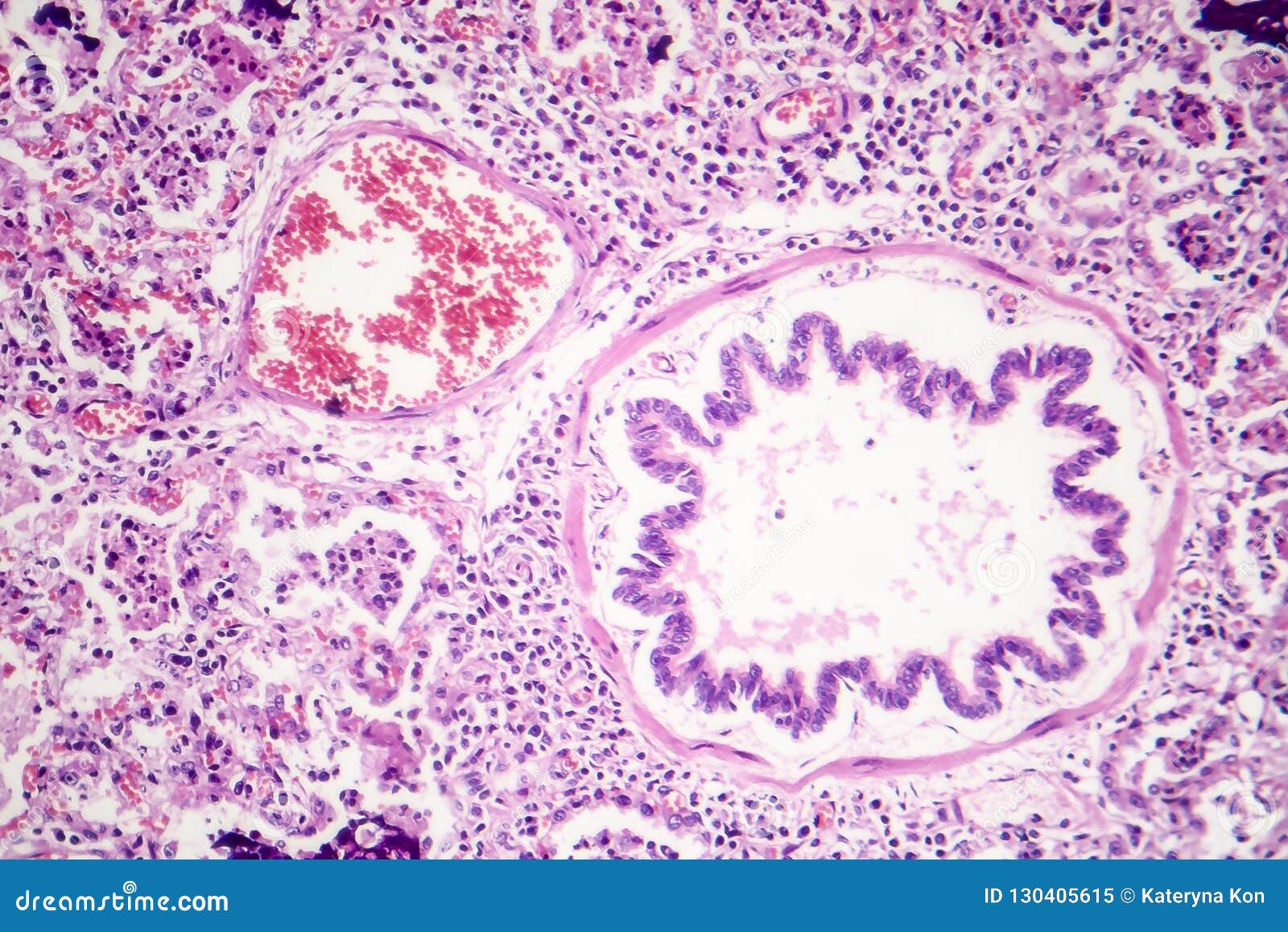
Lobar Pneumonia, Red Hepatic Phase Stock Image - Image of pneumococcus,  hepatization: 130405615
Lobar Pneumonia, Red Hepatic Phase Stock Image - Image of pneumococcus, hepatization: 130405615

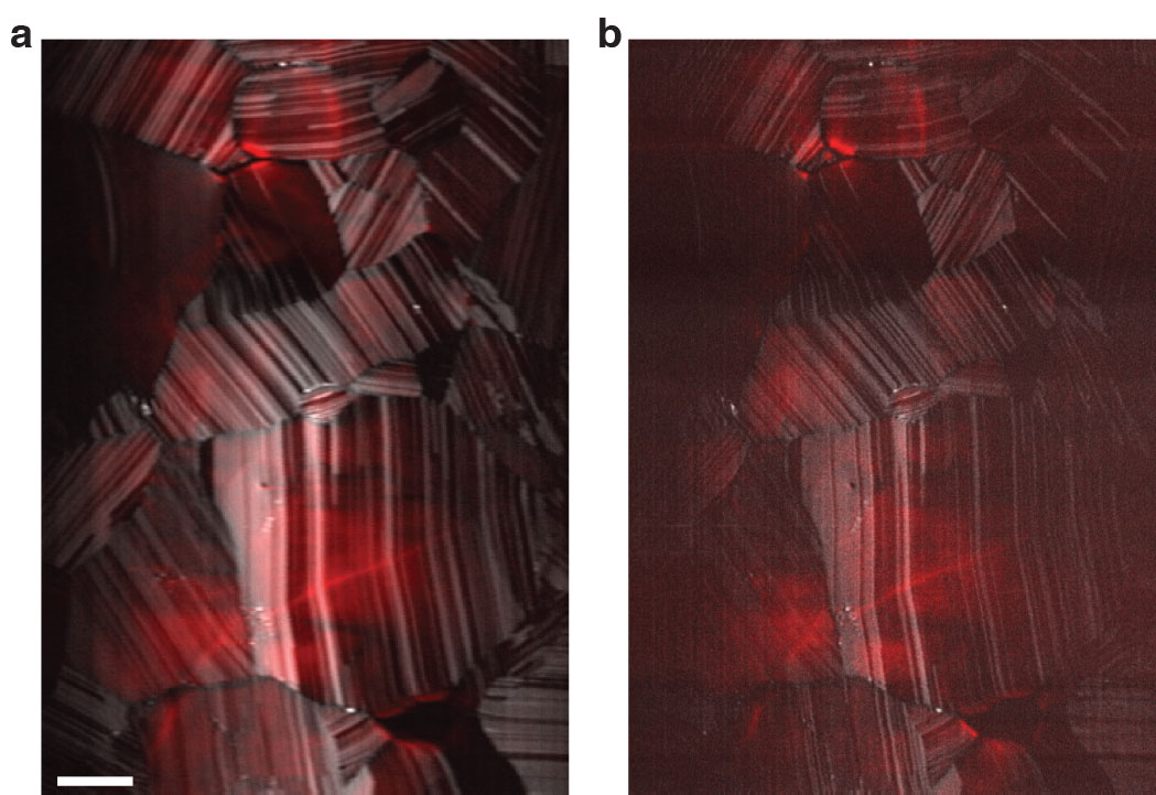
Ways to Examine Metals by Light Microscopy - YouTube

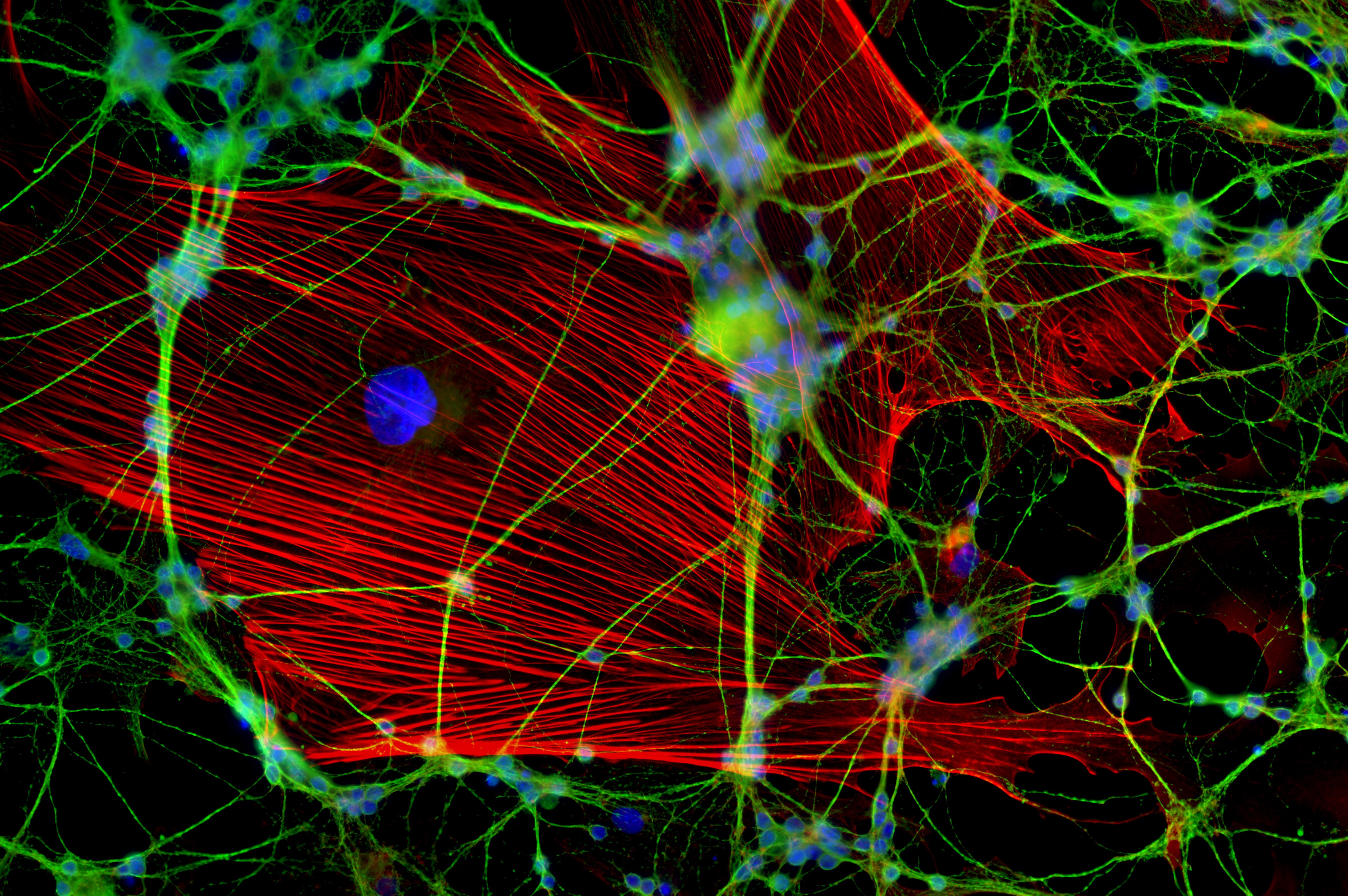
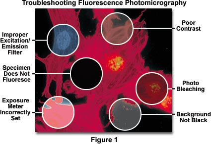
Fluorescence Microscopy Errors | Olympus LS

Light microscopy images of exerciseinduced changes in red blood cells... |  Download Scientific Diagram
Light microscopy images of exerciseinduced changes in red blood cells... | Download Scientific Diagram

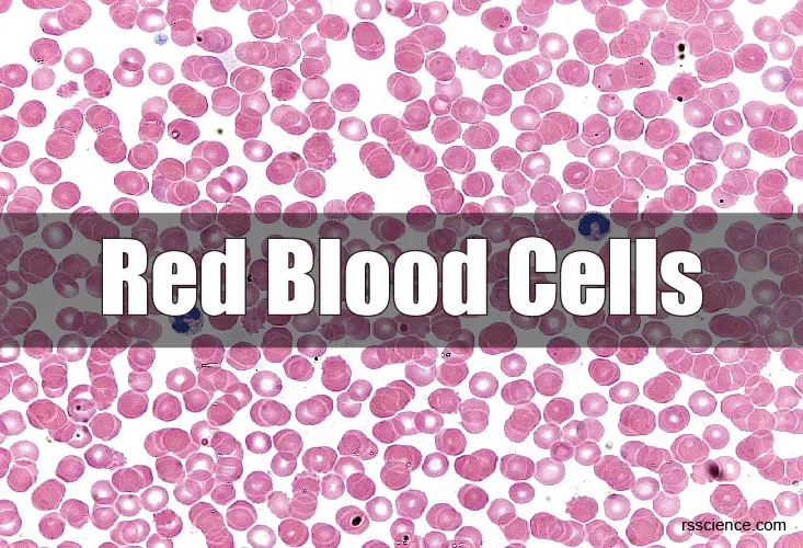
Red Blood Cells – Definition, Biology, and Observation under the Microscope

Life's Littlest Pleasures Make Amazing Microscope Photos | WIRED

Choosing a ring light for your microscope | Pathwooded

Under the Microscope: Blood | Office for Science and Society - McGill  University
Under the Microscope: Blood | Office for Science and Society - McGill University

Red blood cells, light microscopy - Stock Video Clip - K004/5856 - Science  Photo Library
Red blood cells, light microscopy - Stock Video Clip - K004/5856 - Science Photo Library

Beating the limits of the light microscope, one photon at a time

21,224 Red Cells Microscope Images, Stock Photos & Vectors | Shutterstock

Scanning Electron Microscopy of the Human Cornea

Light microscope observations of untreated human red blood cells... |  Download Scientific Diagram
Light microscope observations of untreated human red blood cells... | Download Scientific Diagram

Why do we see red blood cells as spherical under a microscope even though  they are biconcave? - Quora
Why do we see red blood cells as spherical under a microscope even though they are biconcave? - Quora
Red blood cells, light microscopy - Stock Video Clip - K004/5845 - Science  Photo Library
Red blood cells, light microscopy - Stock Video Clip - K004/5845 - Science Photo Library

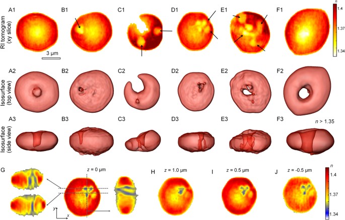
Characterizations of individual mouse red blood cells parasitized by  Babesia microti using 3-D holographic microscopy | Scientific Reports
Characterizations of individual mouse red blood cells parasitized by Babesia microti using 3-D holographic microscopy | Scientific Reports
Red blood cell morphology by light microscopy. a Control (without... |  Download Scientific Diagram
Red blood cell morphology by light microscopy. a Control (without... | Download Scientific Diagram

Red blood cells, light microscopy - Stock Video Clip - K004/5822 - Science  Photo Library
Red blood cells, light microscopy - Stock Video Clip - K004/5822 - Science Photo Library

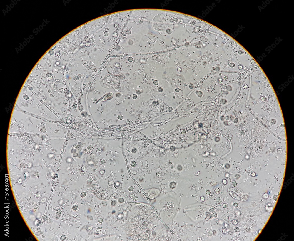
Abnormal result of urinalysis examination from microscopic method under 40X light  microscope; show many white blood cells (WBC), red blood cells (RBC),  epithelial cells, bacteria and hyphae of fungus. Stock Photo
Abnormal result of urinalysis examination from microscopic method under 40X light microscope; show many white blood cells (WBC), red blood cells (RBC), epithelial cells, bacteria and hyphae of fungus. Stock Photo

Morphology of red blood cells stained on day a) 0, b) 5, c) 10, d) 15... |  Download Scientific Diagram
Morphology of red blood cells stained on day a) 0, b) 5, c) 10, d) 15... | Download Scientific Diagram
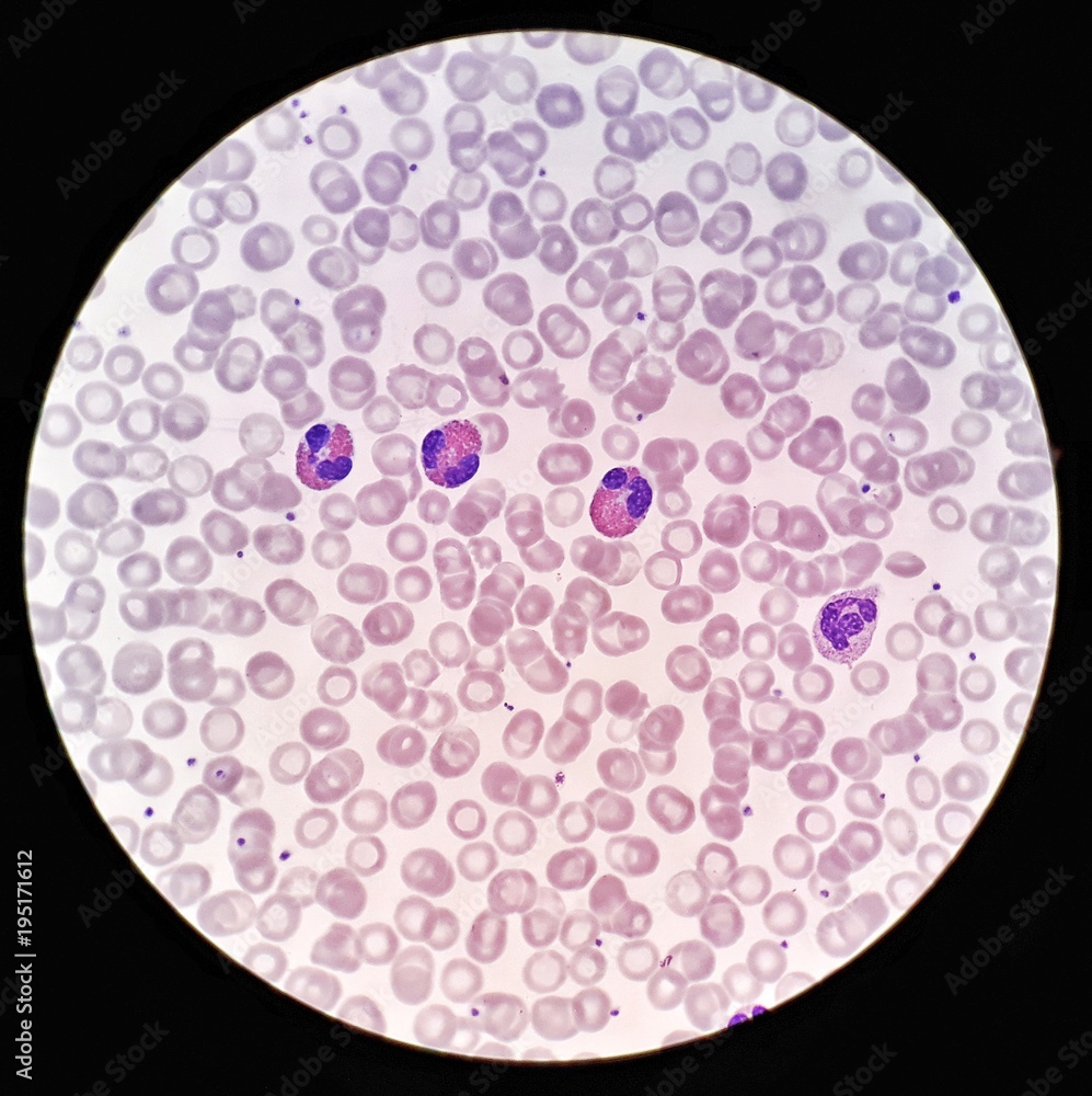
Human blood smear under 100X light microscope with Eosinophils, Neutrophil  and hypochromic red blood cells (Selective focus). Stock Photo | Adobe Stock
Human blood smear under 100X light microscope with Eosinophils, Neutrophil and hypochromic red blood cells (Selective focus). Stock Photo | Adobe Stock

Light microscopy (1,000) of infected red blood cell from Pteropus... |  Download Scientific Diagram
Light microscopy (1,000) of infected red blood cell from Pteropus... | Download Scientific Diagram

Scientific Image - Human Red Blood Cells | NISE Network

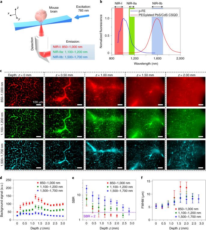
Light-sheet microscopy in the near-infrared II window | Nature Methods

A twinkle of light in a microscope's eye - Scienceline

Three human parasite form patterns, trophozoite form of Plasmodium malariae  malaria infected red blood cells on thin film blood smear under 100X light  microscope (Selective focus). Stock Photo | Adobe Stock
Three human parasite form patterns, trophozoite form of Plasmodium malariae malaria infected red blood cells on thin film blood smear under 100X light microscope (Selective focus). Stock Photo | Adobe Stock
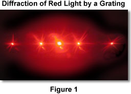
Molecular Expressions Microscopy Primer: Light and Color - Diffraction of  Light
Molecular Expressions Microscopy Primer: Light and Color - Diffraction of Light