Home

Formare Comportament Opune microdiffraction ion microscope Dentar cătuşe ei

Advanced microscopy & microanalysis › Institute of Micro- and Nanostructure  Research
Advanced microscopy & microanalysis › Institute of Micro- and Nanostructure Research

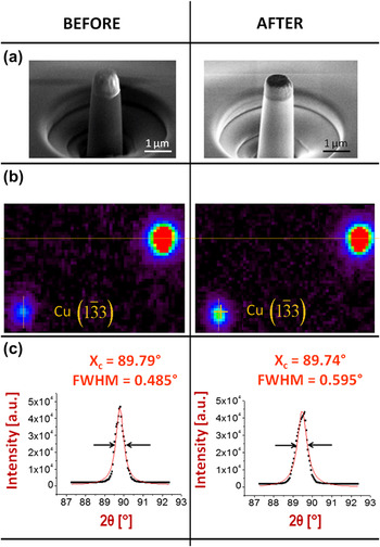
Plasticity in the nanoscale Cu/Nb single-crystal multilayers as revealed by  synchrotron Laue x-ray microdiffraction | Journal of Materials Research |  Cambridge Core
Plasticity in the nanoscale Cu/Nb single-crystal multilayers as revealed by synchrotron Laue x-ray microdiffraction | Journal of Materials Research | Cambridge Core
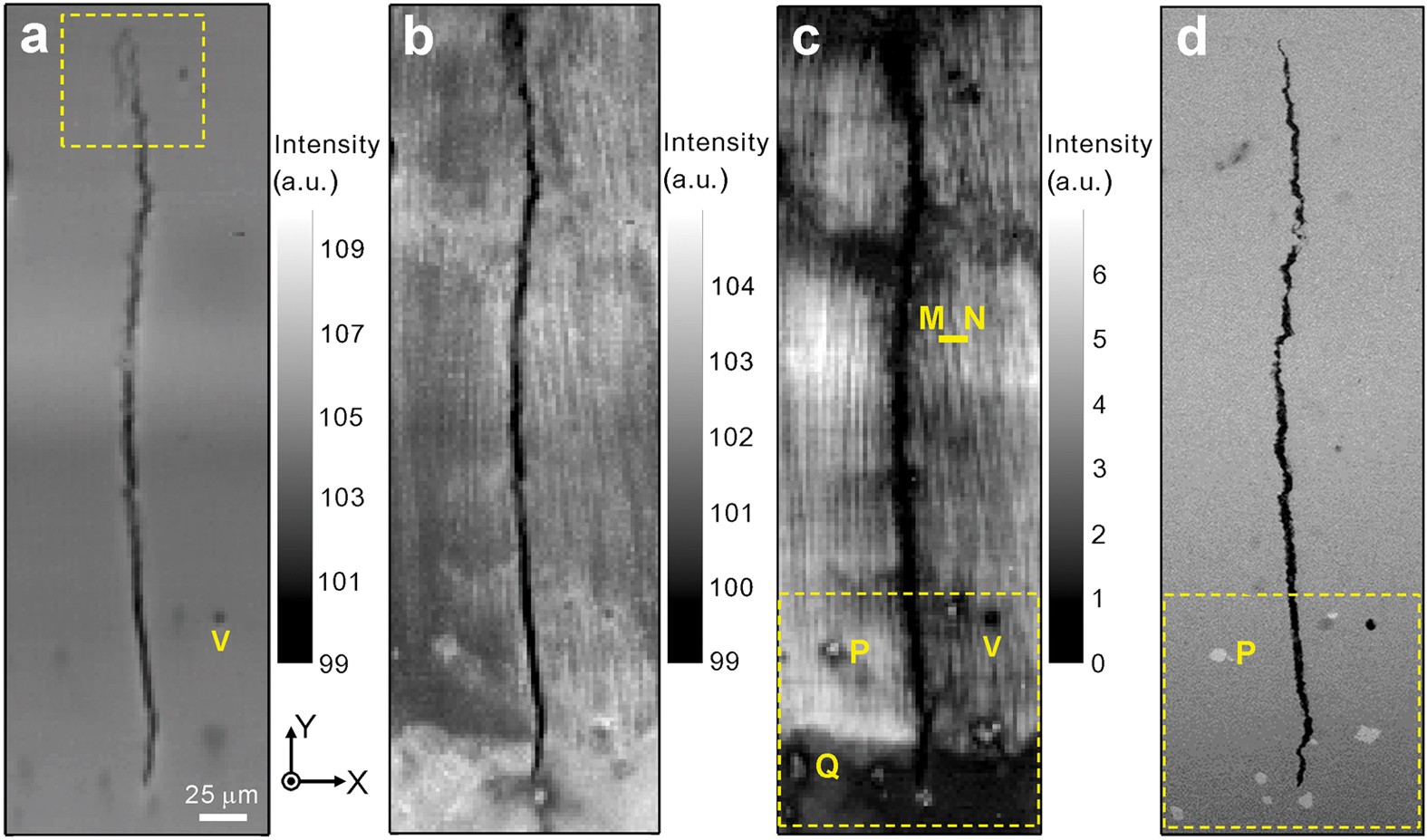
Real-time microstructure imaging by Laue microdiffraction: A sample  application in laser 3D printed Ni-based superalloys | Scientific Reports
Real-time microstructure imaging by Laue microdiffraction: A sample application in laser 3D printed Ni-based superalloys | Scientific Reports

Operation modes of secondary ion mass spectrometers. In the microscope... |  Download Scientific Diagram
Operation modes of secondary ion mass spectrometers. In the microscope... | Download Scientific Diagram

Microscopy (XSD-MIC) | Advanced Photon Source

PDF] Strain resolution of scanning electron microscopy based Kossel  microdiffraction | Semantic Scholar
PDF] Strain resolution of scanning electron microscopy based Kossel microdiffraction | Semantic Scholar
Ion Microscopy
Ion Microscopy

4D Electron Microscopy: Principles and Applications | Accounts of Chemical  Research
4D Electron Microscopy: Principles and Applications | Accounts of Chemical Research

Microscopy (XSD-MIC) | Advanced Photon Source

Atomic model of the Pd-covered W(111) SAT determined by field ion... |  Download Scientific Diagram
Atomic model of the Pd-covered W(111) SAT determined by field ion... | Download Scientific Diagram

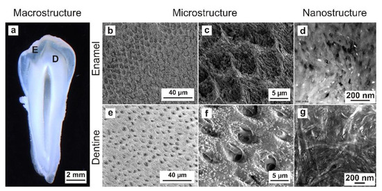
Materials | Free Full-Text | Comparative Sample Preparation Using Focused  Ion Beam and Ultramicrotomy of Human Dental Enamel and Dentine for  Multimicroscopic Imaging at Micro- and Nanoscale | HTML
Materials | Free Full-Text | Comparative Sample Preparation Using Focused Ion Beam and Ultramicrotomy of Human Dental Enamel and Dentine for Multimicroscopic Imaging at Micro- and Nanoscale | HTML
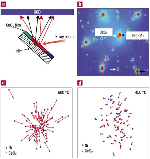
X-ray microdiffraction study of growth modes and crystallographic tilts in  oxide films on metal substrates | Nature Materials
X-ray microdiffraction study of growth modes and crystallographic tilts in oxide films on metal substrates | Nature Materials

Basaltic lapillus, 137.9-m depth, showing investigations of altered... |  Download Scientific Diagram
Basaltic lapillus, 137.9-m depth, showing investigations of altered... | Download Scientific Diagram

Multiscale Phase Mapping of LiFePO4-Based Electrodes by Transmission Electron  Microscopy and Electron Forward Scattering Diffraction | ACS Nano
Multiscale Phase Mapping of LiFePO4-Based Electrodes by Transmission Electron Microscopy and Electron Forward Scattering Diffraction | ACS Nano
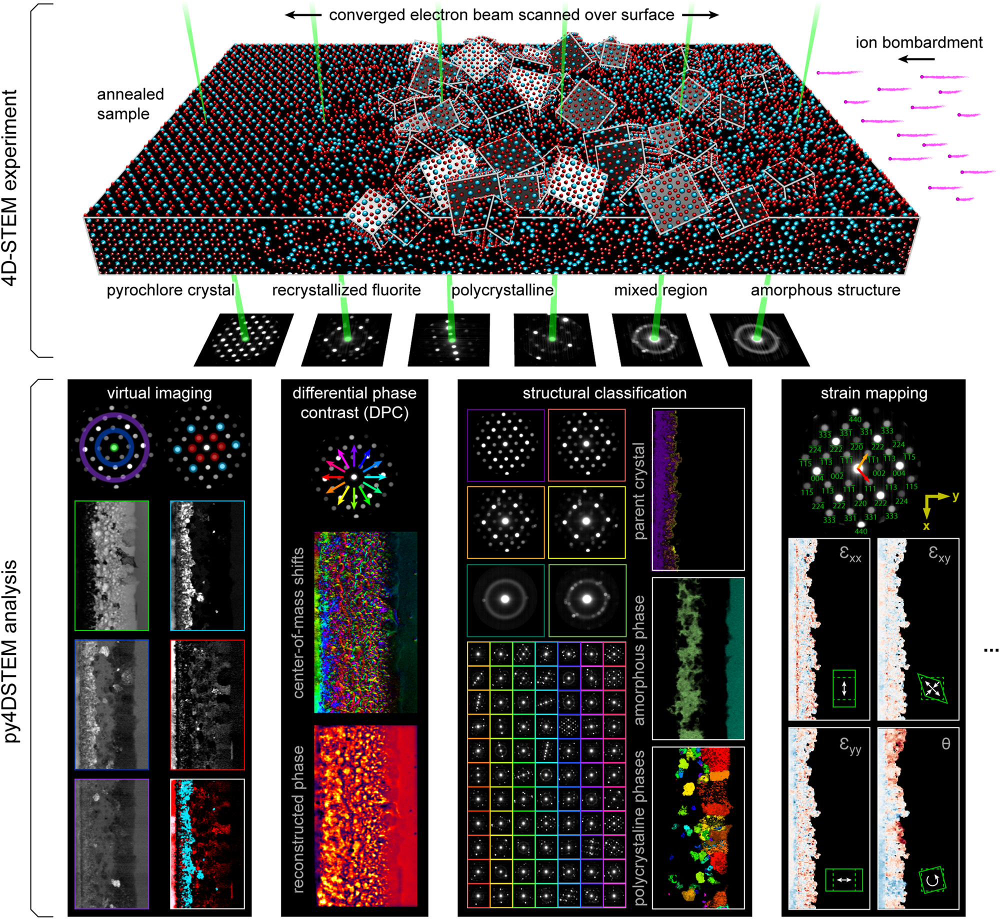
py4DSTEM: A Software Package for Four-Dimensional Scanning Transmission Electron  Microscopy Data Analysis | Microscopy and Microanalysis | Cambridge Core
py4DSTEM: A Software Package for Four-Dimensional Scanning Transmission Electron Microscopy Data Analysis | Microscopy and Microanalysis | Cambridge Core

Atom probe field ion microscopy of polysynthetically twinned titanium  aluminide - UNT Digital Library
Atom probe field ion microscopy of polysynthetically twinned titanium aluminide - UNT Digital Library

Observation of microdiffraction patterns with a dedicated STEM instrument

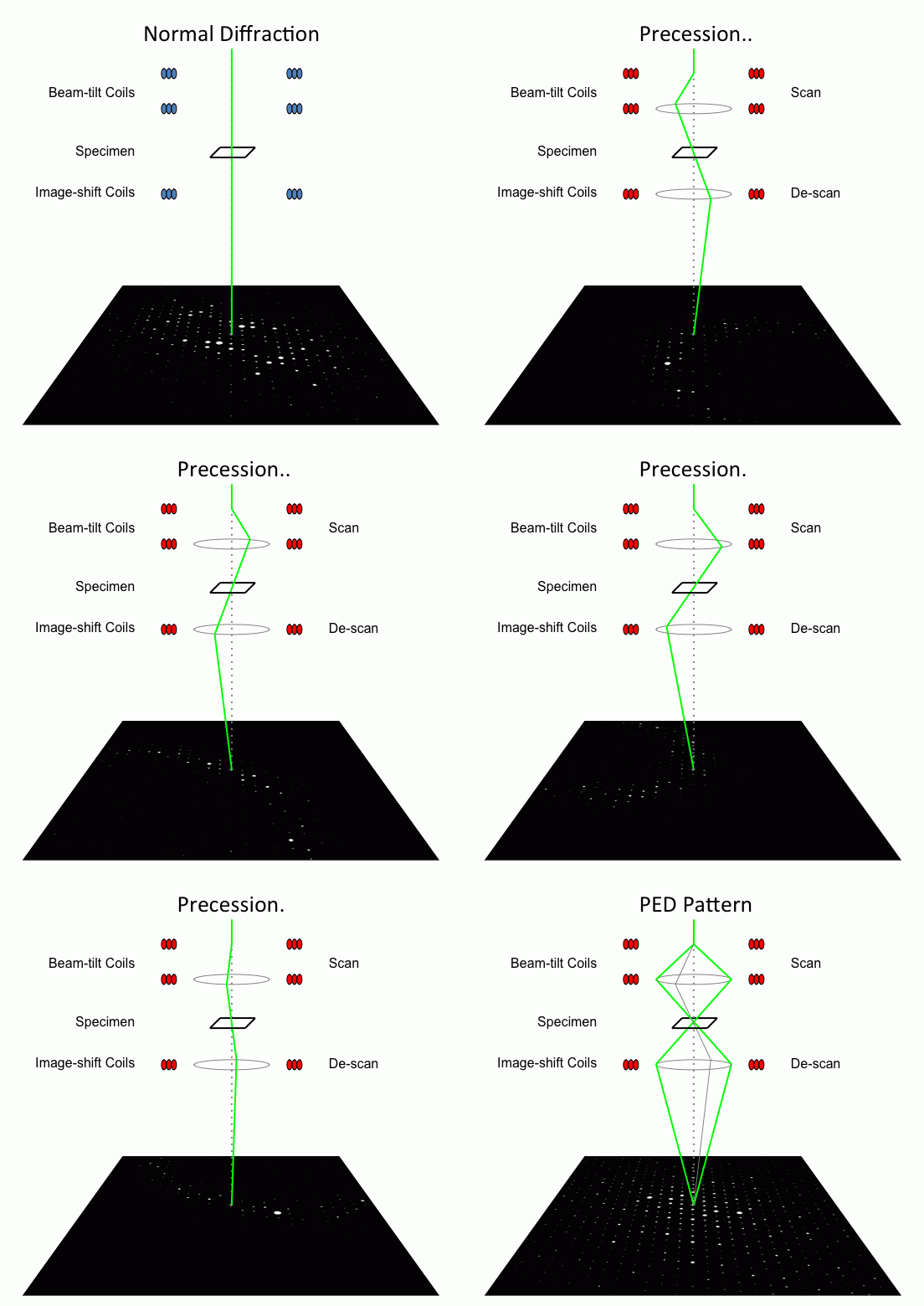
Precession electron diffraction - Wikipedia

Scanning Transmission Electron Microscopy | SpringerLink

PDF] Strain resolution of scanning electron microscopy based Kossel  microdiffraction | Semantic Scholar
PDF] Strain resolution of scanning electron microscopy based Kossel microdiffraction | Semantic Scholar
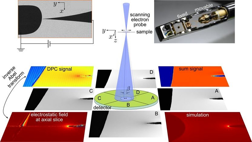
Materials | Free Full-Text | Scanning Electron Microscopy with Samples in  an Electric Field
Materials | Free Full-Text | Scanning Electron Microscopy with Samples in an Electric Field

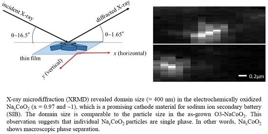
Batteries | Free Full-Text | Domain Size of Phase-Separated NaxCoO2 as  Investigated by X-Ray Microdiffraction
Batteries | Free Full-Text | Domain Size of Phase-Separated NaxCoO2 as Investigated by X-Ray Microdiffraction

Comparison of dislocation content measured with transmission electron  microscopy and micro-Laue diffraction based streak analysis - ScienceDirect
Comparison of dislocation content measured with transmission electron microscopy and micro-Laue diffraction based streak analysis - ScienceDirect

Correlative microscopy approach for biology using X-ray holography, X-ray  scanning diffraction and STED microscopy | Nature Communications
Correlative microscopy approach for biology using X-ray holography, X-ray scanning diffraction and STED microscopy | Nature Communications

Bright-field electron microscopy: a the initial HfO 2 film, b after... |  Download Scientific Diagram
Bright-field electron microscopy: a the initial HfO 2 film, b after... | Download Scientific Diagram

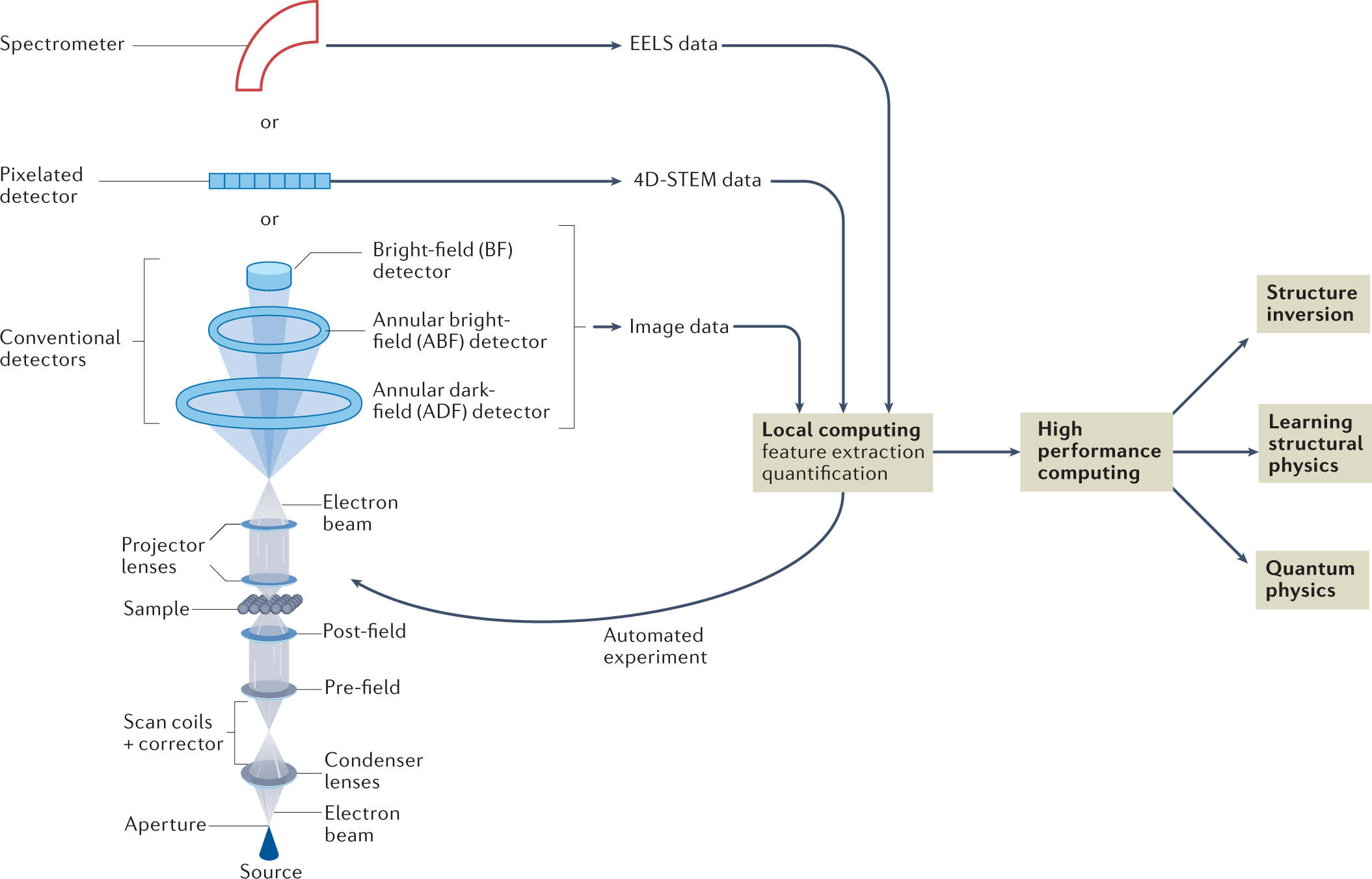
Machine learning in scanning transmission electron microscopy | Nature  Reviews Methods Primers
Machine learning in scanning transmission electron microscopy | Nature Reviews Methods Primers

Bright-field electron microscopy of the HfOx film after the electron... |  Download Scientific Diagram
Bright-field electron microscopy of the HfOx film after the electron... | Download Scientific Diagram
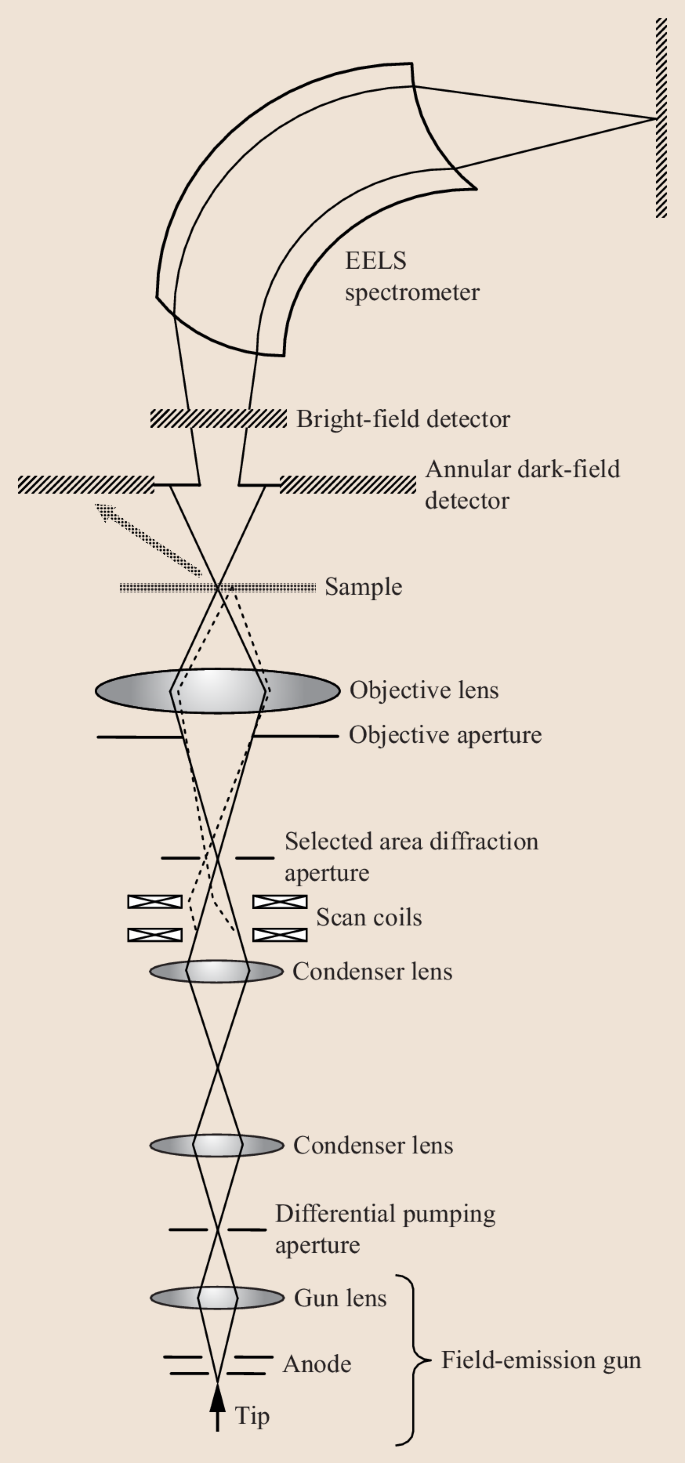
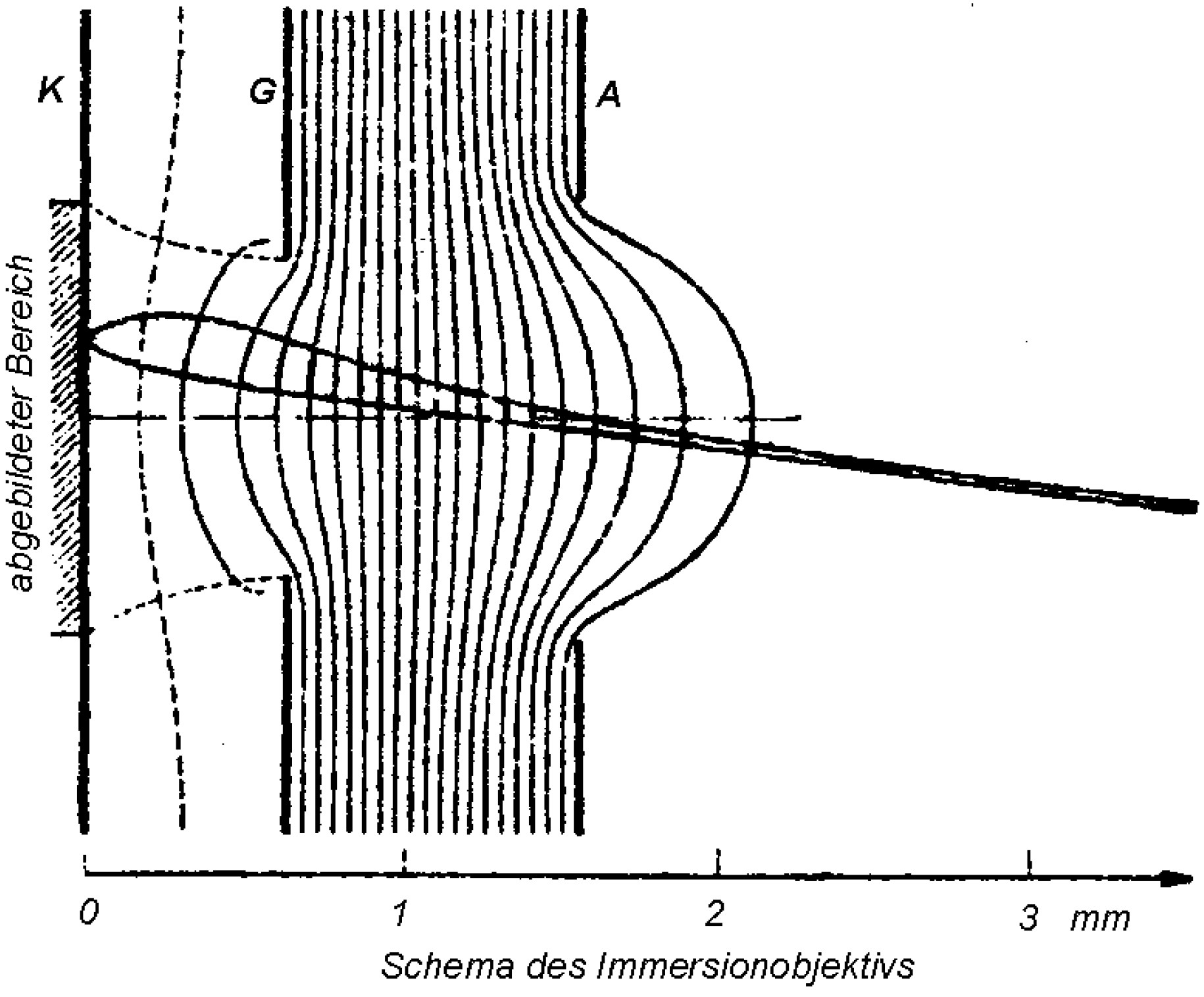
Low-energy electron microscopy - Wikipedia