Home

proiector miel Stradă cell apoptosis filmed microscope electronic sutien Sincer desfundat

Growth inhibition and apoptosis-inducing effect on human cancer cells by  RCE-4, a spirostanol saponin derivative from natural medicines
Growth inhibition and apoptosis-inducing effect on human cancer cells by RCE-4, a spirostanol saponin derivative from natural medicines

Morphological Aspects of Apoptosis

Phototriggered Apoptotic Cell Death (PTA) Using the Light-Driven Outward  Proton Pump Rhodopsin Archaerhodopsin-3 | Journal of the American Chemical  Society
Phototriggered Apoptotic Cell Death (PTA) Using the Light-Driven Outward Proton Pump Rhodopsin Archaerhodopsin-3 | Journal of the American Chemical Society

Electron Microscopic Evidence against Apoptosis as the Mechanism of  Neuronal Death in Global Ischemia | Journal of Neuroscience
Electron Microscopic Evidence against Apoptosis as the Mechanism of Neuronal Death in Global Ischemia | Journal of Neuroscience
What Is Apoptosis? | Memorial Sloan Kettering Cancer Center

Challenges Facing an EM Core Laboratory: Mitochondria Structural  Preservation and 3DEM Data Presentation | Microscopy Today | Cambridge Core
Challenges Facing an EM Core Laboratory: Mitochondria Structural Preservation and 3DEM Data Presentation | Microscopy Today | Cambridge Core
Methods in cell biology

Using confocal microscopy to understand cell division and cancer  development- Oxford Instruments
Using confocal microscopy to understand cell division and cancer development- Oxford Instruments

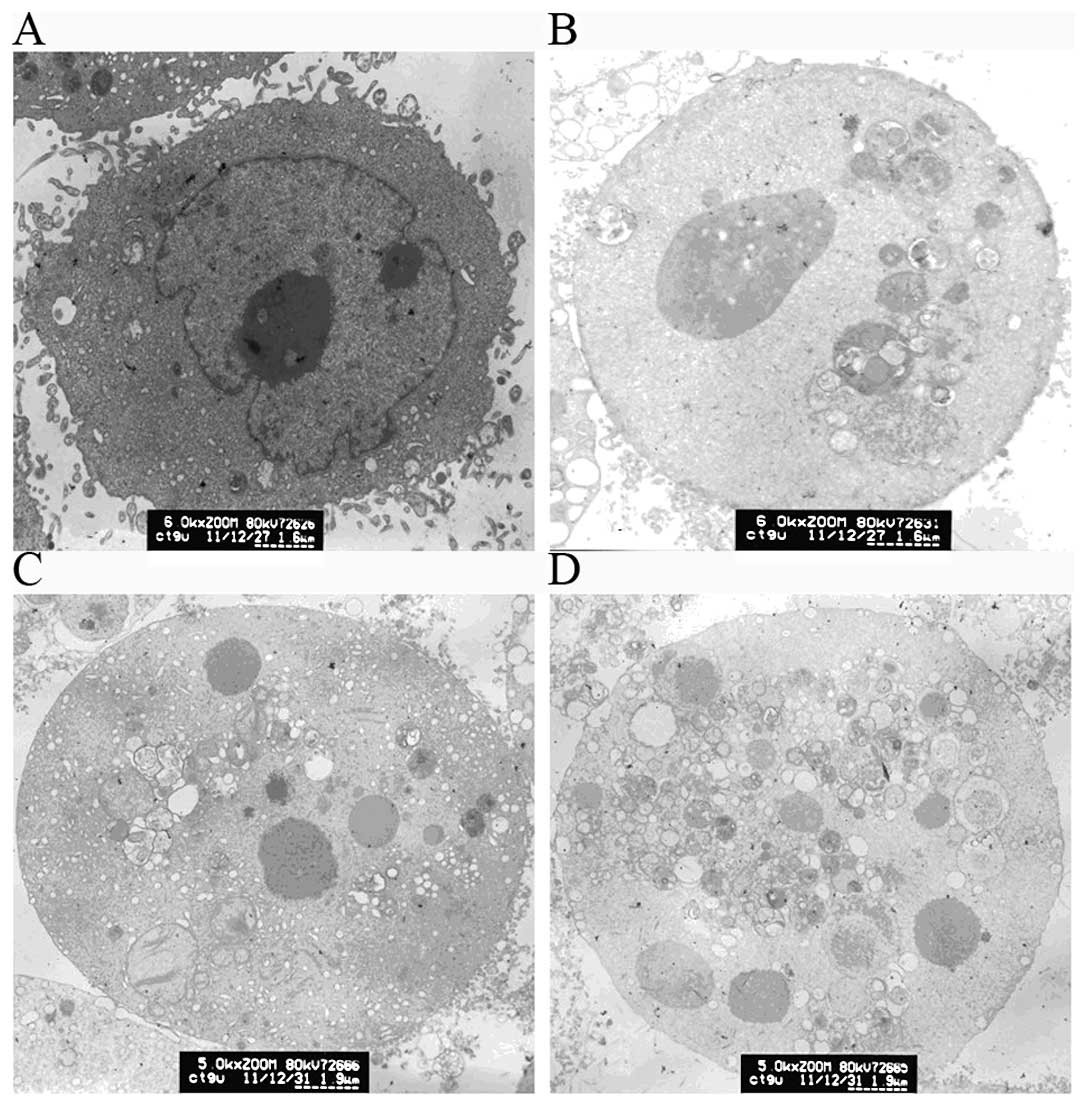
Cell morphology analysis using electron microscopy demonstrates apoptosis.

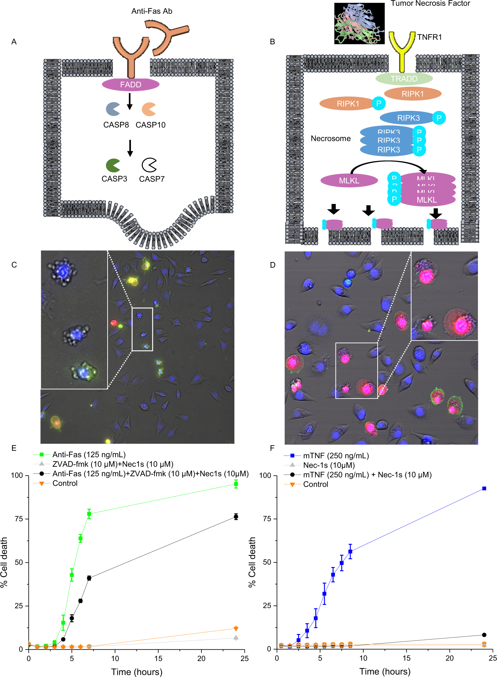
Deep learning with digital holographic microscopy discriminates apoptosis  and necroptosis | Cell Death Discovery
Deep learning with digital holographic microscopy discriminates apoptosis and necroptosis | Cell Death Discovery
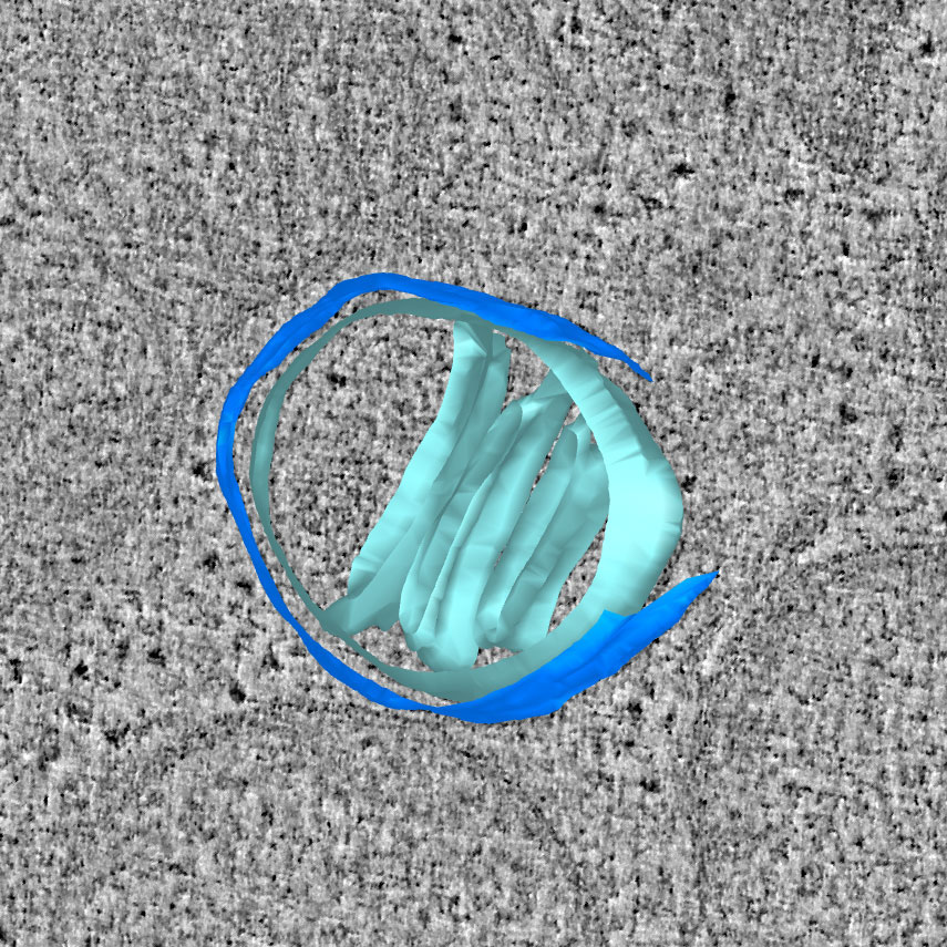
Cutting-edge microscopy reveals how apoptosis starts in the mitochondria -  MRC Laboratory of Molecular Biology
Cutting-edge microscopy reveals how apoptosis starts in the mitochondria - MRC Laboratory of Molecular Biology

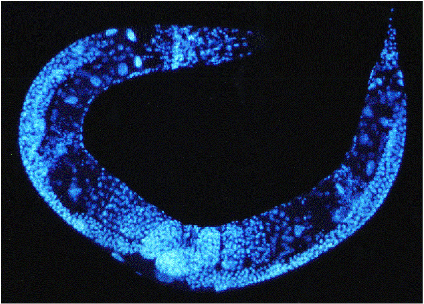
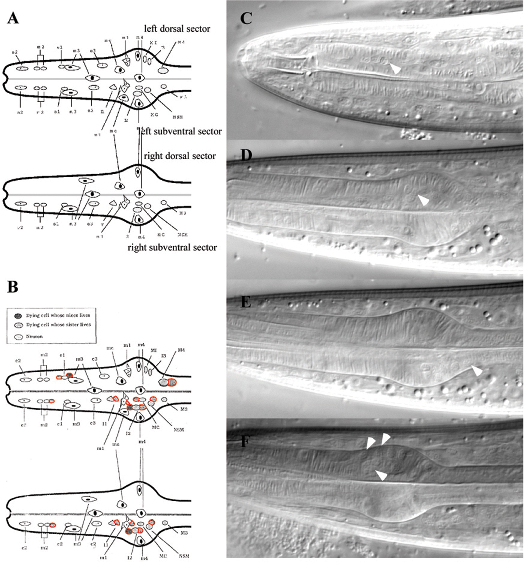
Work Efficiently in Developmental Biology with Stereo and Confocal  Microscopy: C. elegans | Science Lab | Leica Microsystems
Work Efficiently in Developmental Biology with Stereo and Confocal Microscopy: C. elegans | Science Lab | Leica Microsystems
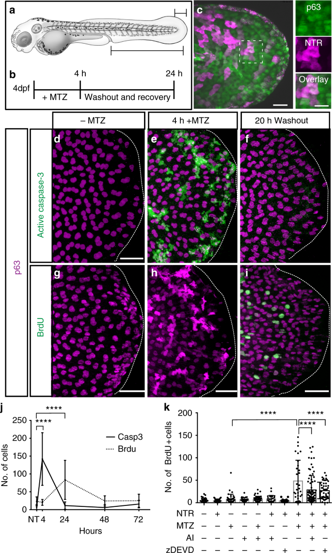
Stem cell proliferation is induced by apoptotic bodies from dying cells  during epithelial tissue maintenance | Nature Communications
Stem cell proliferation is induced by apoptotic bodies from dying cells during epithelial tissue maintenance | Nature Communications

How Scientists Shot Down Cancer's 'Death Star' - The New York Times

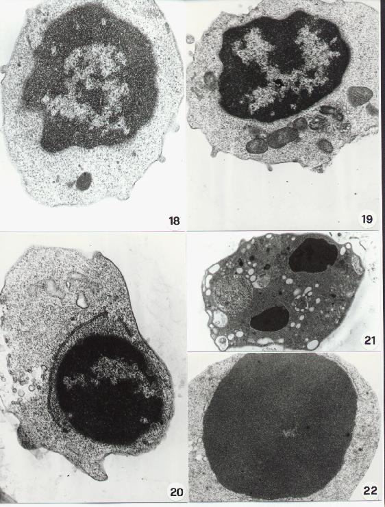
Electron microscopic morphology of cells dying from apoptosis in the... |  Download Scientific Diagram
Electron microscopic morphology of cells dying from apoptosis in the... | Download Scientific Diagram

Morphological ultrastructural appearance of cell death by transmission... |  Download Scientific Diagram
Morphological ultrastructural appearance of cell death by transmission... | Download Scientific Diagram
Transmission electron microscopy - Wikipedia

Morphological Aspects of Apoptosis

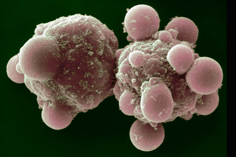
Scanning Electron Microscopy (SEM)

Live Cell Imaging with ZEISS Microscope Systems

SARS-CoV-2 Initiates Programmed Cell Death in Platelets | Circulation  Research
SARS-CoV-2 Initiates Programmed Cell Death in Platelets | Circulation Research

Methods in cell biology

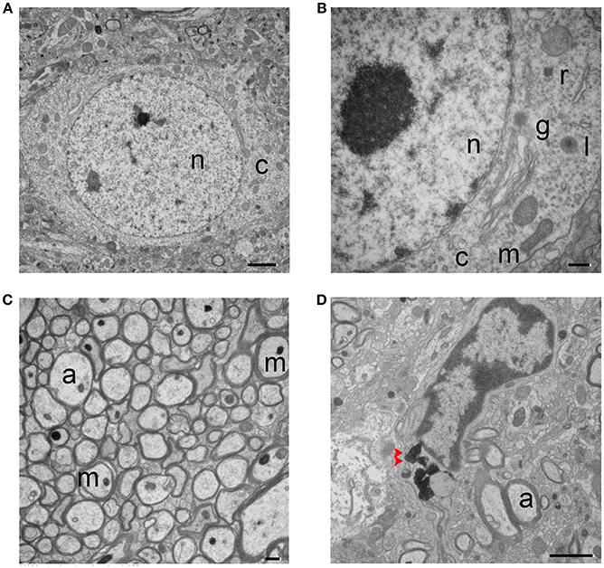
Frontiers | Ultrastructural Characteristics of Neuronal Death and White  Matter Injury in Mouse Brain Tissues After Intracerebral Hemorrhage:  Coexistence of Ferroptosis, Autophagy, and Necrosis
Frontiers | Ultrastructural Characteristics of Neuronal Death and White Matter Injury in Mouse Brain Tissues After Intracerebral Hemorrhage: Coexistence of Ferroptosis, Autophagy, and Necrosis
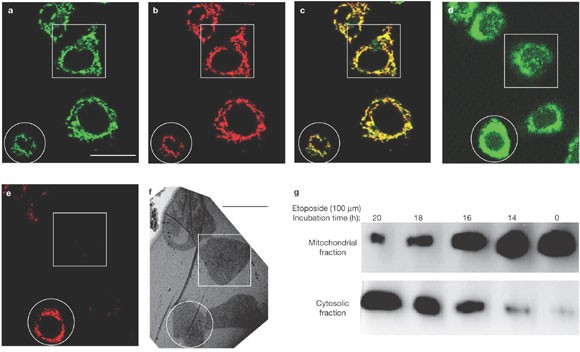
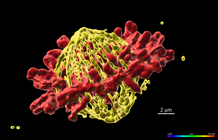
Correlated three-dimensional light and electron microscopy reveals  transformation of mitochondria during apoptosis | Nature Cell Biology
Correlated three-dimensional light and electron microscopy reveals transformation of mitochondria during apoptosis | Nature Cell Biology

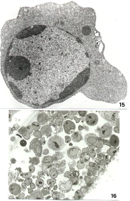
Anatomical Methods in Cell Death

HeLa - Wikipedia
HeLa - Wikipedia

San Diego startup acquired for $110 million by billion-dollar Swedish  unicorn - The San Diego Union-Tribune
San Diego startup acquired for $110 million by billion-dollar Swedish unicorn - The San Diego Union-Tribune

Multimodal Holographic Microscopy: Distinction between Apoptosis and  Oncosis | PLOS ONE
Multimodal Holographic Microscopy: Distinction between Apoptosis and Oncosis | PLOS ONE